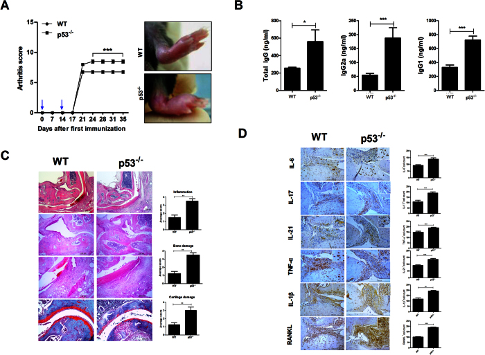
Figure 5. p53 deficiency exacerbates CIA severity.
Mice were sacrificed on day 35 after the first immunization. (A) Clinical scores ankle condition in CIA induced WT and p53−/− mice (***P < 0.05, n = 10). (B) The expression levels of IgG, IgG1, and IgG2a antibodies in each group were examined. Data are presented as mean ± SD of three independent experiments (*P < 0.05, ***P < 0.01, n = 10). (C) Joint tissues from CIA induced WT and p53−/− mice after staining with H&E (original magnification, 40× or 200×, n = 6) or safranin O (original magnification, 200×, n = 6). (D) Immunohistochemical detection of IL-6, IL-21, IL-17, IL-1β, TNF-α, and RANKL in the synovium of CIA induced WT or p53−/− mice after staining (original magnification, 200×, n = 6). All histological analyses were performed at least 3 times. Representative images are revealed. Data are presented as mean ± SD of three independent experiments (**P < 0.03, ***P < 0.01).

