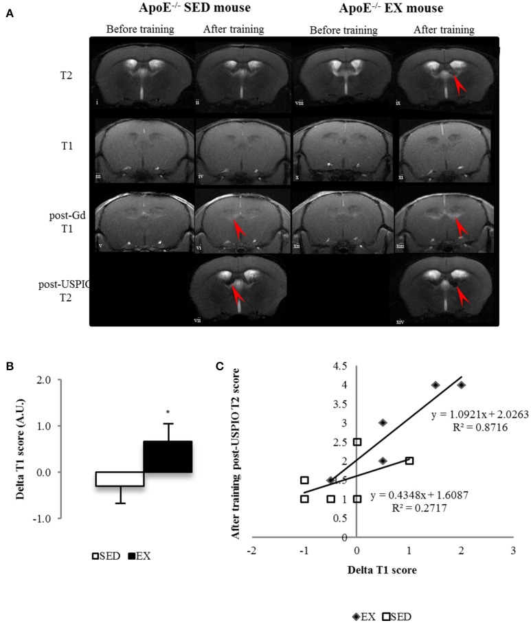
Figure 3.
Longitudinal brain MR images of a SED (i–vii) and an EX ApoE−/− mouse (viii–xiv). before (i, viii) and after training (ii, ix) native T2 image; native (iii, iv, x, xi) and Post-Gadolinium (Gd) T1 images (v, vi, xii, xiii), the enhancing bright zone after Gd showing BBB leakage in ApoE−/− mouse (vi, red arrow); Post-USPIO (vii, xiv) T2 images, the loss of signal showing USPIO phagocytosis by macrophages in ApoE−/− (vii, red arrow) (A) Delta score of longitudinal brain MR Imaging before and after-training score of post-Gd T1 (B) in SED and EX ApoE−/− mice (B) (p = 0.02 compared to SED). Linear regression of Delta T1 (ΔT1) scores and after-training post-USPIOs T2 scores of EX and SED ApoE−/− mice showed a stronger correlation between increased BBB leakage and high phagocytic activity in EX mice (C) (p = 0.049). Significantly different from SED mice: *p < 0.05.

