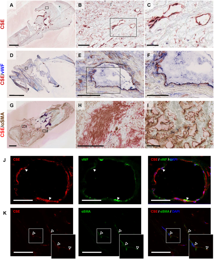
Figure 3. CSE is present in microvascular endothelial cells and αSMA+ cells.
Representative photomicrographs of a plaque with CSE expression in intraplaque microvessels (A–C). Immunohistochemical double staining for CSE and vWF immunohistochemical staining, (D) showing double positivity in microvascular endothelial cells (E,F). CSE protein is visualized with AEC (red) and vWF with FastBlue (blue). Immunohistochemical double staining for CSE and αSMA (G), showing double positivity in intraplaque smooth muscle cells (H), but only CSE positivity in intraplaque microvessels (I). CSE protein is visualized with FastRed (red) and αSMA with DAB (brown). Scalebar: 2 mm (A,D,G); 100 μm (B,E,H); and 50 μm (C,F,I). Immunofluorescence double staining for CSE and vWF, showing co-localization in microvascular endothelial cells (closed arrowheads), 400× magnification, scalebar: 20 μm (J). Immunofluorescence double staining for CSE and αSMA, showing co-localization of CSE and αSMA in myofibroblast-like cells (open arrowheads), 400× magnification, scalebar: 20 μm (K).

