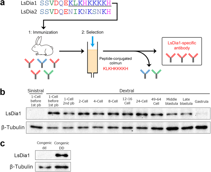
Figure 3. Generation of an LsDia1-specific antibody and developmental expression of LsDia1 protein.
(a) Comparison of the amino acid sequences of the C-terminal region between LsDia1 (residues 1054–1068) and LsDia2 (residues 1066–1080), which were selected as a target site for making anti-LsDia1 antibody. Underlined sequence indicates the recognition site of anti-LsDia1 polyclonal antibodies. (b) Western blotting analyses showed that LsDia1 protein is present in the dextral embryos from 1-cell immediately after oviposition to the blastula stage, but is not detectable for the sinistral embryos even at the 1-cell stage prior to 1st pb extrusion. LsDia1 and β-Tubulin levels were analyzed sequentially using the same blot. The whole images of blot showing the single band of correct MW for LsDia1 and β-Tubulin, respectively, are presented in Supplementary Figure S4. (c) Presence and absence of LsDia1 protein in early stage embryos of the congenic (F10) dd strain (1–4-cell) and DD strain (1-cell), respectively.

