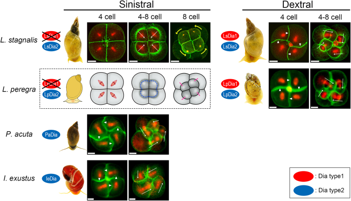
Figure 4. Dia gene types and SD/SI at the third cleavages for the three pond snails studied.
Fixed embryos during the third cleavage were stained for actin filament (green) and β-tubulin (red). White arrowheads and arrows indicate SD and orientation of spindles, respectively. Yellow arrows indicate the rotating direction of small blastomeres towards the 8-cell stage of sinistral L. stagnalis embryo. All were observed from the animal pole side. Scale bar: 20 μm. Sinistral only snails, P. acuta and I. exustus, possess only Dia2 type proteins (blue) (PaDia and IeDia), and exhibit counter-clockwise SD and SI. Snails which belong to Lymnaeidae, L. stagnalis and L. peregra have both dominant dextral and recessive sinistral animals in the wild. The dominant dextral ones of the two species have both Dia type 1 (red) and Dia type 2 (blue) proteins and exhibit clockwise SD and SI, whereas the recessive sinistral L. stagnalis possesses only Dia type 2 (blue) protein showing no SD/SI. Figures for L. stagnalis were adapted from Shibazaki et al.21. Lack of SD/SI was observed for sinistral L. peregra, but no photographs of the adult snail nor embryo images are available, and hence are shown schematically. (Sinistral L. peregra is now unavailable as a laboratory strain). The photographs of snails were taken by ourselves.

