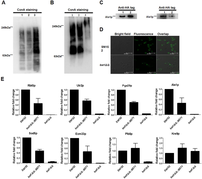
Figure 2. Cell wall anchorage of GPI-APs in BST1-deficient C. albicans strains was seriously impaired.
(A) Intracellular GPI-APs from crude protein extracts prepared from exponentially growing parent strain (SN152) (Lane 1), BST1-complemented (bst1Δ/Δ::BST1) (Lane 2) and bst1Δ/Δ mutant strains (Lane 3) underwent immunoblotting using peroxidase labeled-ConA. (B) HF-released cell wall anchored GPI-Aps from cell wall debris of exponentially growing parent strain (SN152) (Lane 1), BST1-complemented (bst1Δ/Δ::BST1) (Lane 2) and bst1Δ/Δ mutant strains (Lane 3) underwent immunoblotting using peroxidase labeled-ConA. (C) Als1p extracted from crude protein extracts (left panel) or HF-treatment cell wall debris (right panel) of parent (als1Δ/ALS1-HA) (Lane 1), bst1Δ/Δ mutant strain (bst1Δ/Δ als1Δ/ALS1-HA) (Lane 2) using anti-HA tag antibody. (D) Fluorescence images exhibiting cell wall anchorage of Als1p in parent and bst1Δ/Δ mutant strains by confocal laser scanning microscope using anti-HA tag antibody. (E) Relative fold change of representative and virulence related GPI-APs with impaired cell wall anchorage in BST1-deficient C. albicans strains. The cell wall proteins of SN152, bst1Δ/Δ::BST1, and bst1Δ/Δ strains were analyzed by LC-MS/MS on high-resolution instruments (LTQ-Orbitrap XL and Velos, Thermo Fisher). Raw files were processed by MaxQuant (version 1.3.0.5) for peptide/protein identification and quantification.

