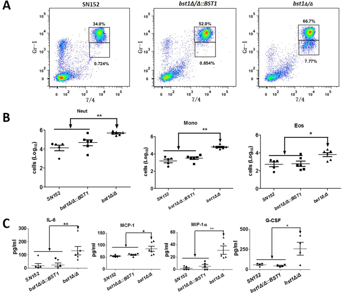
Figure 6. Enhanced in vivo inflammatory response to C. albicans by bst1 deficiency.
(A) Flow cytometry for Gr-1hi7/4hi neutrophils and Gr-1+ 7/4hi inflammatory monocytes in mice 4 hours following intraperitoneal infection with 5 × 10⁵ UV-inactivated yeast phase parent (SN152), BST1-complemented (bst1Δ/Δ::BST1), bst1Δ/Δ null mutant strains. Numbers adjacent to outlined areas indicate the percentage of neutrophils (top panel) and monocytes (lower panel). Data are representative images of 5 mice. (B) Scatter plots of myeloid cell subsets in the peritoneal cavities of mice after 4 hours of intraperitoneal infection with 5 × 10⁵ UV-inactivated yeast phase parent (SN152), BST1-complemented (bst1Δ/Δ::BST1), bst1Δ/Δ null mutant strains. Each symbol represents an individual mouse. Data are representative of three independent experiments.*P < 0.05; **P < 0.01 (Kruskal-Wallis nonparametric One-way ANOVA with Dunns post-test). (C) ELISA for cytokines, chemokines and growth factors in lavage fluid from the inflamed peritoneal cavities of mice after 4 hours of intraperitoneal infection with 5 × 10⁵ UV-inactivated yeast phase parent (SN152), BST1-complemented (bst1Δ/Δ::BST1), bst1Δ/Δ null mutant strains. IL-6, MCP-1, MIP-1α, G-CSF, GM-CSF. Data are representative of three independent experiments. *P < 0.05; **P < 0.01 (Kruskal-Wallis nonparametric One-way ANOVA with Dunns post-test).

