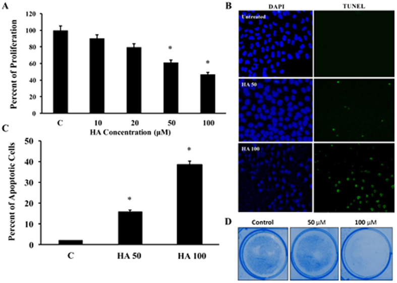
Figure 1.
(A) Hep G2.2.1.5 cells were cultured with different concentrations of HA (10, 20, 50 and 100 μM) for 24 hours and the cell proliferation assay was performed by MTT assay to check the cell viability (n = 3; *p < 0.05). (B) Apoptosis induction by HA was measured in Hep G2.2.1.5 cells by incubating with 50 and 100 μM of HA using TUNEL assay and the representative picture from 3 experiments is shown. (C) The total number of positive cells were counted in at least 10 different high power fields and the average is presented. (n = 3; *p < 0.05). (D) The colony formation assay was performed in 60 mm dishes with 50 or 100 μM HA (n = 3).

