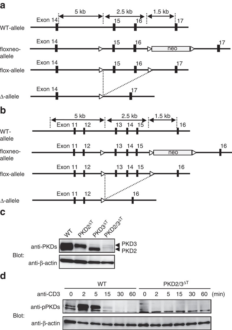
Figure 2. Generation of PKD-deficient mice.
(a,b) Genomic structures and targeting constructs of PKD2 (a) and PKD3 (b). Exons encoding the DxxxN motif and the phosphorylation sites required for kinase activity are flanked by loxP sites (indicated by open triangles). (c) Total thymocytes from WT, PKD2fl/fl × Lck-Cre Tg (PKD2ΔT), PKD3fl/fl × Lck-Cre Tg (PKD3ΔT) and PKD2fl/fl × PKD3fl/fl × Lck-Cre Tg (PKD2/3ΔT) mice were analysed for PKD expression by western blotting using anti-PKDs antibody. β-actin was used as a loading control. (d) PKD phosphorylation in thymocytes on TCR stimulation by CD3 cross-linking for 2 min was detected using anti-pPKDs. Data are representative of two independent experiments (c,d).

