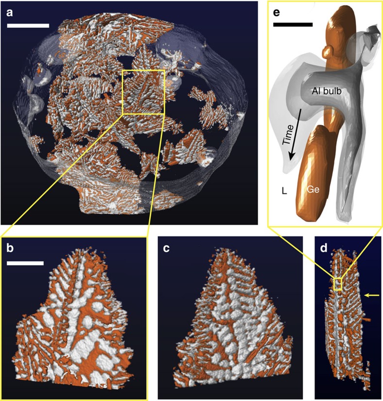
Figure 2. Irregular eutectic microstructures across length scales.
(a) Snapshot of the full tomographic region-of-interest during the growth process, where Ge is orange, Al is white and the melt is blue. One eutectic colony is boxed and isolated for subsequent 3D imaging. (b–d) The front (0°), back (180°), and side (90°) views of this eutectic colony. The arrow in d points to a region of high structural regularity, wherein the Ge lamellae are nearly parallel. The front and back views (b,c) suggest that pockets of Al cover the exposed Ge surfaces. (e) In some cases, defects cause holes or gaps within the Ge plates; then, the Al bulbs can grow through these holes and spread across Ge {111}. Scale bar, 200 μm (a), 70 μm (b) and 10 μm (e).

