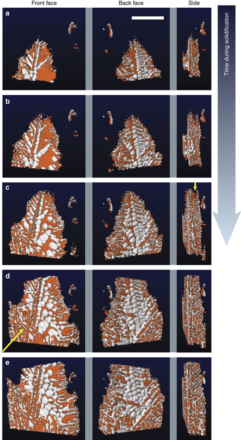
Figure 3. Morphology of the eutectic colony during the growth process.
Reconstructions given at (a) 100, (b) 140, (c) 180, (d) 220 and (e) 260 s after the start of solidification. Shown are three views per time-step, corresponding to the front (0°), back (180°) and side (90°) of the eutectic colony. The arrow in the side view of c points to the tips of the Ge plates that lead the solidification event; the arrow in the front view of d points to another eutectic colony that impinges upon the one of interest. When viewed from either the front or the back, the interfacial morphology is markedly different from that predicted by Fisher and Kurz1: bulbous-like domains of Al envelope the surfaces of Ge as solidification proceeds. Scale bar, 100 μm.

