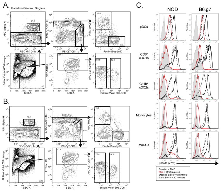
Fig. 5. Phosphoflow for monocytic and DC populations.
Splenocytes were harvested and pooled from 8–10 week old NOD and B6.g7 mice for signaling experiments. The gating scheme for the fixed and permeabilized cells isolated from (A) NOD mice and (B) B6.g7 mice. (C) Splenocytes were stimulated in vitro with 10 ng/ml IFN-gamma for 5 min (dashed black) and 30 min (solid black), and then stained intracellularly for pSTAT1. Unstimulated cells are represented by solid red line. Fluorescence minus one is represented by solid gray histogram. The data shown is representative of three separate experiments.

