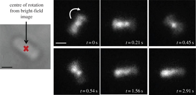
Figure 4.
Frames from video fluorescence microscopy performed over 3 s showing single molecules diffusing/binding inside a tethered rotating electrocompetent E. coli RP437, FliCst cell electroporated with 1.25 µM, i.e. 25 pmol, CheY(Cys)-Cy3B: at times during the movie (e.g. at t = 0 s, t = 0.21 s and t = 0.45 s), it is possible to clearly see a bright spot on top of the centre of rotation, indicating a possible interaction of CheY with the motor; at other times (e.g. at t = 1.56 s and t = 2.91 s), the bright spot localizes at one pole of the cell; finally, in some frames (e.g. at t = 0.21 s and t = 0.54 s), it is possible to observe two bright spots in the cell, one of which is always at the centre of rotation and the other seems to be located at—or travelling to—the cell pole. Exposure time 30 ms, intensity used for Cy3B: 400 nW µm−2; intensity used for Atto647: 0.86 µW µm−2. Scale bar = 1 µm. (Substack of full video given in the electronic supplementary material, CheYmot1 movie (20 fps).)

