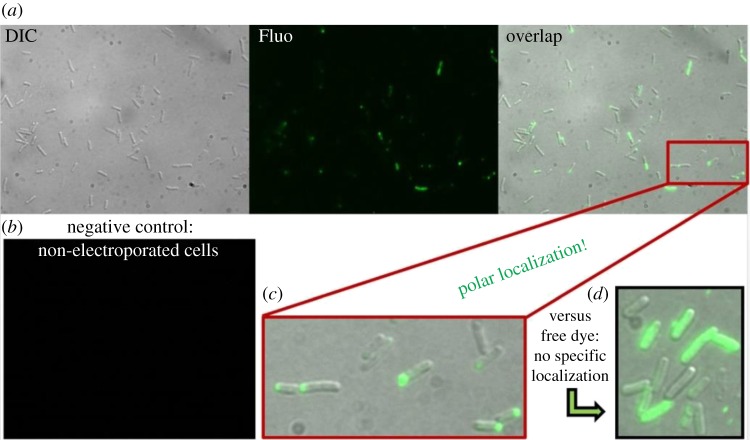
Figure 6.
(a) Fluorescence images in false colours of electrocompetent E. coli cells electroporated with 1.5 µM (30 pmol) CheY(Cys)-Cy3B imaged in DIC, fluorescence and an overlap of the two; (b) electrocompetent cells incubated with the same amount of protein but not electroporated showed little or no fluorescence internalization. (c) Detail showing polar localization of electroporated CheY-Cy3B proteins inside the cells; (d) Further control: electroporated free Cy3B-maleimide dye shows no specific localization. Epifluorescence, 150 ms exposure time. Electroporation parameters and procedure as described in §2c.

