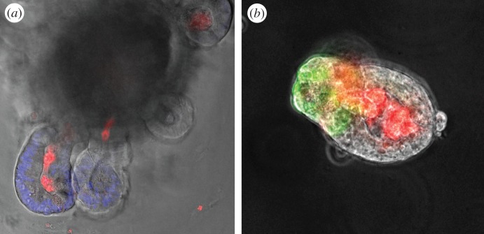
Figure 2.
Typical images of intestinal organoids treated with MDP-rhodamine, showing that the MDP is internalized in the lumen of the organoid during the embedding process. In (a) organoid from wild-type mouse (nuclei are stained in blue with DAPI and MDP in red) and in (b) from lgr5-EGFP mouse (stem cells are in green and MDP in red).

