
Fig. 2.
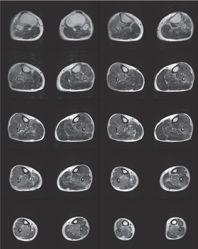
T2-weighted magnetic resonance imaging of the lower extremities in the late phase. The anterior, lateral, and deep posterior compartments of both calf muscles are swollen, and the signal intensity is increased. Inner compartment enhancement is not observed, but the fascia rim is enhanced. The superficial posterior compartment shows mild swelling, and signal intensity is increased heterogeneously. There is patchy enhancement in the superficial posterior compartment.