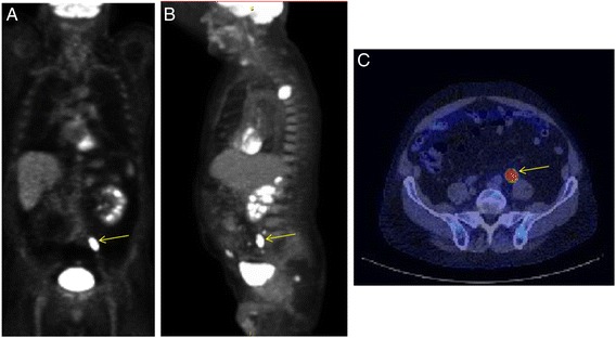
Fig. 2.

Focal [18 F]FDG uptake in a 79 year old male who was evaluated for an isolated right upper lobe pulmonary nodule. (a) coronal and (b) sagittal PET slices show indeterminate FDG avid foci (arrows) within the rectum. Area of increased uptake (arrow) was localized by PET/CT (c). Tissue biopsy revealed invasive moderately differentiated (grade 3 of 4) adenocarcinoma
