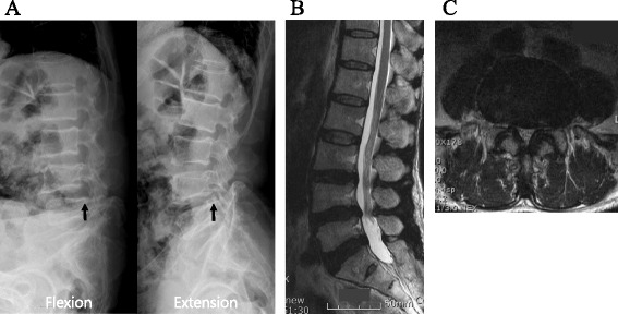
Fig. 1.

Preoperative radiographs and lumbar magnetic resonance imaging. a Dynamic plain radiographs showing degenerative spondylolisthesis of lumbar vertebral body 4 on lumbar vertebral body 5. The radiographic finding showed anterior slipping of lumbar vertebral body 4 on lumbar vertebral body 5 when the patient bent his back (arrow). An extension view shows the slight reduction of anterior slippage of lumbar vertebral body 4 on lumbar vertebral body 5 (arrow). b Sagittal view showing disc protrusion and stenosis of both foramen on lumbar vertebral body 4 and lumbar vertebral body 5. The dural sac was compressed ventrally and dorsally at lumbar vertebral body 4 and lumbar vertebral body 5 on a sagittal view of T2-weighted images. c Axial view showing disc protrusion, stenosis, and facet hypertrophy on lumbar vertebral body 4 and lumbar vertebral body 5. Narrowing of the spinal canal was found with both disc protrusion and ligamentum flavum hypertrophy
