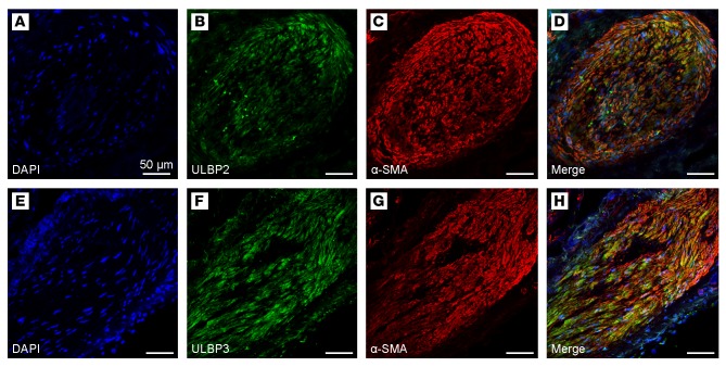
Figure 2. NKG2D ligand an α-SMA localization in lymphangioleiomyomatosis (LAM) cells in lung biopsy specimens from LAM patients.
(A–D and E–H) Immunofluorescent images show nodular LAM lesions from 2 distinct LAM patients. LAM lung tissue sections were stained for DAPI (A and E), the NKG2D ligands (B, green) ULBP2 and (F, green) ULBP3, as well as for the LAM cell marker (C, G, red) α-SMA. Confocal microscopy images show immunofluorescent localization of the NKG2D ligands (D) ULBP2 and (H) ULBP3 with α-SMA within the LAM cells comprising the nodular LAM lesions. Scale bars: 50 μm. The images are representative of results obtained by immunofluorescent staining of diagnostic lung biopsies from 9 LAM patients.

