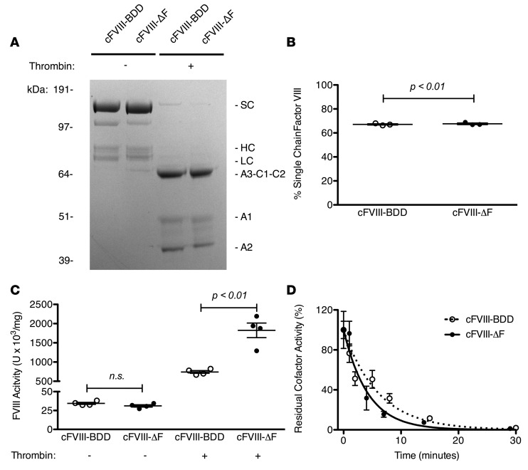
Figure 7. Biochemical characterization of recombinant canine factor VIII-ΔF (cFVIII-ΔF ) compared with cFVIII-BDD.
(A) SDS-PAGE analysis of 3 μg of cFVIII-BDD and cFVIII-ΔF staining with Coomassie blue before (−) or after (+) activation with thrombin. Identified protein species are single chain (SC), heavy chain (HC), and light chain (LC). (B) Quantification of percentage of SC of each cFVIII variant by densitometric analysis of SDS-PAGE. Each data point represents a distinct measurement. (C) cFVIII clotting activity was determined by 1-stage or 2-stage clotting assay. Each data point represents a distinct dilution. (D) Decay of cFVIII variants following thrombin activation. Error bars represent SEM of at least 3 separate dilutions. Lines are single-exponential fittings. The half-lives of activated cFVIII-ΔF and cFVIII-BDD are 2.2 ± 0.2 and 4.7 ± 0.4 minutes, respectively; R2 = 0.99 and 0.99, respectively. Means were compared by 2-tailed Student’s t test. P values greater than 0.05 considered not significant (n.s.). Horizontal markers in whisker plots represent the mean and 1 SEM.

