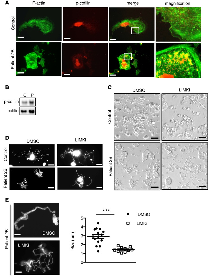
Figure 7. Human type 2B mutant vWF/p.V1316M (2B) megakaryocytes (MKs) exhibit defective actin structure and proplatelet formation, which is rescued by LIM kinase (LIMK) inhibition.
(A) Control and patient 2B MKs after thrombopoietin-induced differentiation in culture were incubated over a fibrinogen matrix for 3 hours and stained for F-actin (green) and the Ser3-phosphorylated cofilin (p-cofilin, red). Scale bars: 10 μm. White squares indicate the magnification zone. (B) Western blot of p-cofilin and total cofilin in control (C) and in patient MKs (P). (C) Proplatelet formation in liquid culture in the presence or the absence of LIMKi at day 11, in control and in patient MKs. LIMKi (10 μM) was added at day 10; DMSO (0.2%) was used as vehicle. Scale bars: 50 μm. (D) Control and patient 2B MKs after thrombopoietin-induced differentiation in culture were spread over a polylysine-coated coverslip for 3 hours and stained for F-actin. Scale bars: 20 μm. (E) Proplatelet structure after F-actin staining and graph of the size of platelet-like structures in the 2B patient in the presence or absence of LIMKi. Scale bars: 5 μm. MKs in the presence of DMSO (n = 15) and MKs in the presence of LIMKi (n = 10) were analyzed for the size of platelet-like structures. Statistical significance was determined by Student’s t test. ***P < 0.001.

