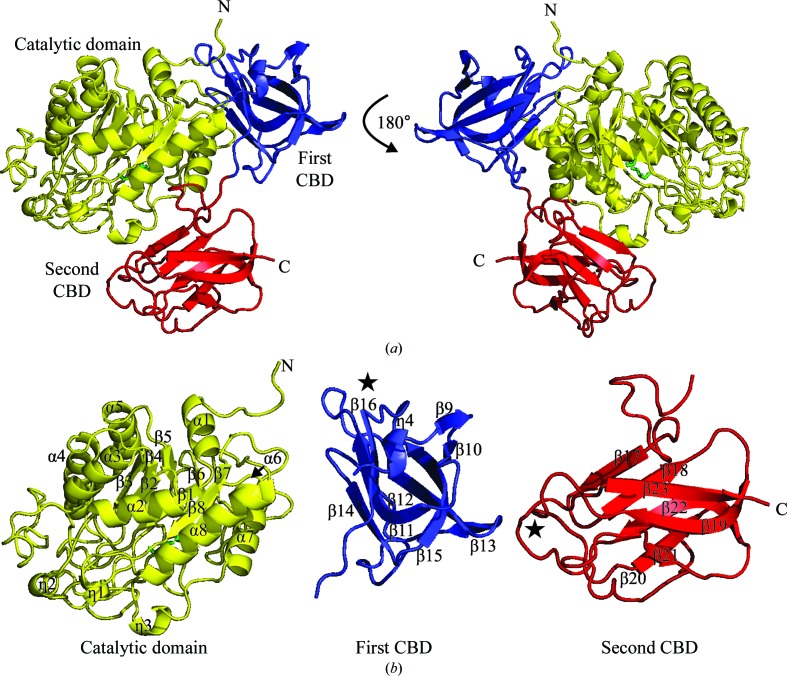
Figure 2.
The structure of GH29_0940. (a) Cartoon representation of the GH29 monomer. The catalytic domain is coloured yellow, the first carbohydrate-binding domain (CBD) blue and the second CBD red. Two glycerol molecules (shown as green sticks) are present in the active site of the catalytic domain. (b) Three-dimensional structure of each domain of GH29_0940 individually. The α-helical segments and β-strands are labelled. The 310-helices are indicated by η. The filled stars indicate the region containing the predicted substrate-binding residues. All figures were produced using PyMOL v.1.3 (http://www.pymol.org; Schrödinger).

