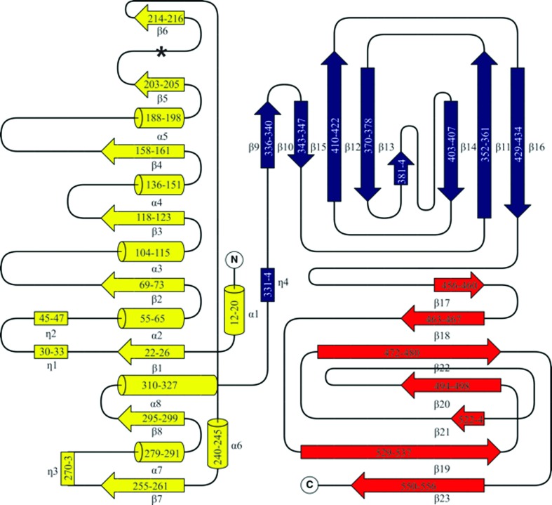
Figure 3.
Topology of GH29_0940. β-Strands are represented by arrows, 310-helices by rectangles and α-helices are shown as cylinders. The topology is coloured according to the domains: the catalytic domain is coloured yellow, the first CBD is in blue and the second CBD is in red. The numbering reflects the residues involved in each secondary structure. The asterisk represents the location of the missing α-helix usually positioned as the fifth helix surrounding the β-barrel (which typically has a total of nine α-helices including the N-terminal α-helix ‘cap’). This figure was drawn with TopDraw (within CCP4) with manual modifications (Bond, 2003 ▸; Winn et al., 2011 ▸).

