
Figure 1.
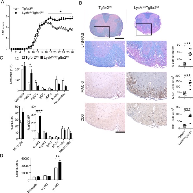
Loss of TGFβ‐signaling in myeloid cells results in chronic EAE without remission. EAE was induced by immunization with MOG emulsified in CFA. Clinical score was assessed on a daily basis (A). Data are pooled from three experiments. n = 25 (Tgfbr2fl/fl), 38 (LysMCreTgfbr2fl/fl). B: Spinal cords taken at day 28 post‐immunization stained with LFB‐PAS, anti‐Mac‐3, and anti‐CD3. n = 7‐8/group. C: Analysis of CNS cell subsets by flow cytometry at day 28 postimmunization n = 3–5/group. moDC monocyte‐derived DC, myDC conventional myeloid DC, lyDC lymphoid DC pDC plasmacytoid DC. D: Surface MHC II expression n = 9/group. Data in A, C, and D are presented as means ± SEM. * P < 0.05 *** P < 0.001 (A, Mann‐Whitney test; B–D, Student's unpaired t‐test). Scale bar = 500 μm (spinal cords) and 200 μm (magnifications). [Color figure can be viewed in the online issue, which is available at wileyonlinelibrary.com.]