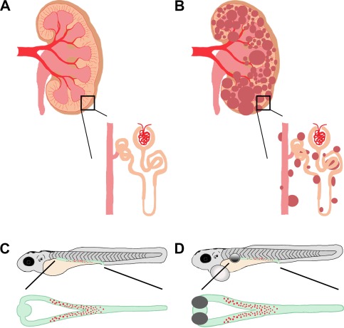
Figure 5.

PKD in mammals and zebrafish. (A, B) Schematic of a healthy, adult mammalian kidney with enlarged nephron (A) compared to a diseased, polycystic adult mammalian kidney and enlarged nephron (B). The collecting duct system is in pink, nephron tubule is in orange, and cysts are in dark red. (C, D) Drawing of zebrafish larvae where the kidney is highlighted in green with red MCCs. (C) A healthy zebrafish larva with an enlarged, dorsal view of a healthy embryonic kidney. (D) Schematic of an unhealthy zebrafish larva, with bilateral kidney cysts (grey circle) and pericardial edema (white circle). Enlarged is a dorsal view of the pronephros, showing the bilateral kidney cysts (grey) as well as distended tubular lumen (green) compared to the wild‐type zebrafish embryonic kidney.
