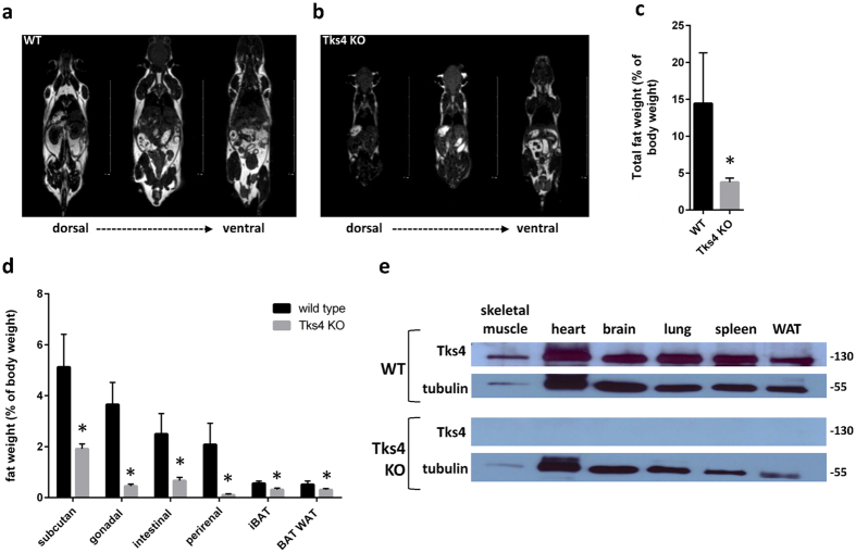
Figure 2. Characterization of Tks4-deficient MSCs.
MRI measurement showing fat tissues (white) and other tissues (gray or black), (a) represents a 7 months old wild type male mouse and (b) represents a 7 months old Tks4 deficient male mouse. (c) Total fat weight measured in three adult WT and Tks4−/− mice. (d) Weights of various fat depos isolated from 7 months old WT and Tks4 KO mice. Three adult mice in each group were analyzed. (e) Skeletal muscle, brain, heart, lung, WAT (gonadal white adipose tissue) and spleen lysates from WT and KO mice were analyzed by Western blot for Tks4. Samples and gels were handled and run under the same experimental conditions. Tubulin was used to control equal loading. *p < 0.05. An unpaired t-test was used to determine the significance of the difference between means of two groups. Error bars represent s.d.

