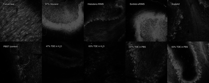
Figure 1.

Human cerebellar cortex stained with anti‐neurofilament primary antibody and subsequently AlexaFluor‐568‐conjugated donkey anti‐mouse secondary antibody and imaged using Leica SP5 confocal microscope with a 10× objective, Z‐projections of Z‐stacks 500‐μm thick (z‐stack step size 5 μm), each imaged using the same settings.
