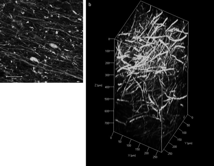
Figure 2.

Immunofluorescence with neurofilament (NF) staining on human cortical tissue. a: A two‐dimensional image of NF staining showing fine axonal processes and neuronal somas. Scale bar = 50 μm. b: Z‐stack image of NF immunostaining on a 3‐mm block of human cortex with an imaging depth to 771.67 μm (z‐stack step size 5.3 μm).
