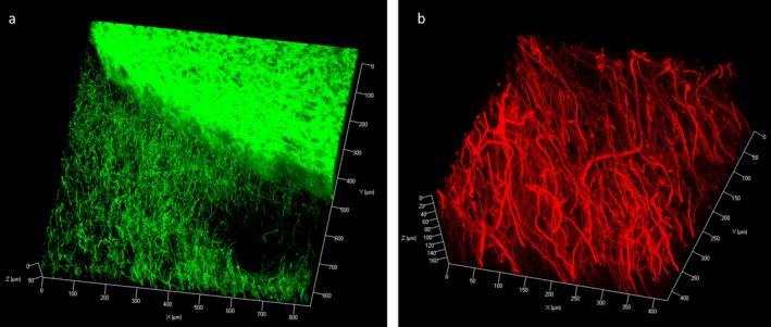
Figure 3.

Z‐stack image of Immunofluorescence with tyrosine hydroxylase (TH) staining on rat coronal block showing TH‐positive neuronal processes at the cortex and dense, homogenous staining within the striatum (z‐stack step size 3.3 μm) (a). Staining in the human midbrain block showed dense TH‐positive axonal processes (z‐stack step size 1.5 μm) (b).
