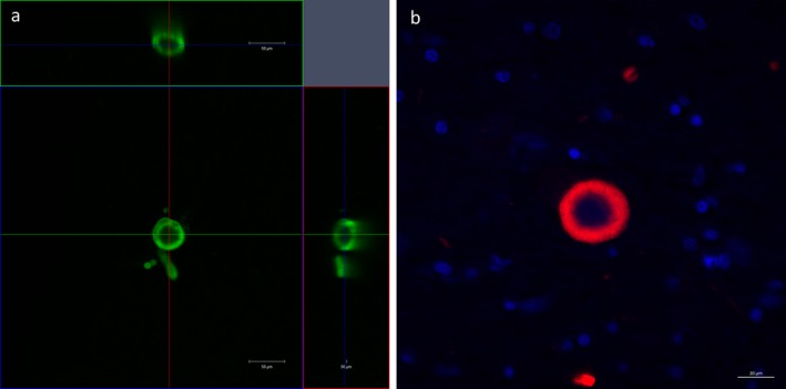
Figure 5.

An orthogonal projection of a Lewy body‐like inclusion in a block of cleared tissue containing the nucleus basalis of Meynert in human stained with anti‐α SN antibody (green), showing a near‐spherical α SN shell of the inclusion in the XY, XZ and YZ planes (Scale bar = 50 μm) (a). A confocal Image of a Lewy body‐like inclusion in a standard 7‐μm‐thick section containing the nucleus basalis of Meynert stained with single immunofluorescence with anti‐α SN antibody (red) and a nuclear counterstain (DAPI, blue) (Scale bar = 20 μm) (b).
