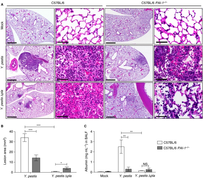
Figure 4.

Plasminogen activator inhibitor‐1 (PAI‐1) enhances lung pathology during Yersinia pestis infection. (A) Sections of formalin‐fixed lungs stained with hematoxylin and eosin from C57BL/6 or C57BL/6 PAI‐1 −/− mice inoculated with phosphate‐buffered saline (mock), 104 colony‐forming units (CFUs) of Y. pestis, or 104 CFUs of Y. pestis ∆pla. Representative images of inflammatory lesions are shown (n = 3). For each infection, the scale bar of the left image (× 2.5 magnification) represents 100 μm, and the scale bar of the right image (× 63 magnification) represents 50 μm. (B) Inflamed lesions were analyzed with imagej to calculate the area of inflammation. Data represent the lesion area (mm2) per field in two sections from three mice, each at × 2.5 magnification. (C) Quantification by ELISA of albumin levels in the bronchoalveolar lavage fluid (BALF) of C57BL/6 or C57BL/6 PAI‐1 −/− mice infected with 104 CFU of Y. pestis or Y. pestis ∆pla, or mock‐infected. Data from two independent experiments are combined (n = 10 for each group), and error bars represent standard errors of the mean (SEMs). One‐way anova with Bonferroni's multiple comparison test was used to determine significance (*P ≤ 0.05; **P ≤ 0.01; ***P ≤ 0.001). NS, not significant.
