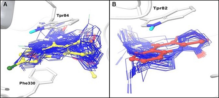
Figure 2.

Predicted binding conformations of new ligands. The most populated clusters are showed in stick representation (blue). Reference ligands, huprine (yellow) and tacrine (red) are shown in ball & stick representation. The binding site residues are shown in stick representation (white). (A) acetylcholinesterase and (B) butyrylcholinesterase.
