. 2016 Sep 23;19(3):231–241. doi: 10.4048/jbc.2016.19.3.231

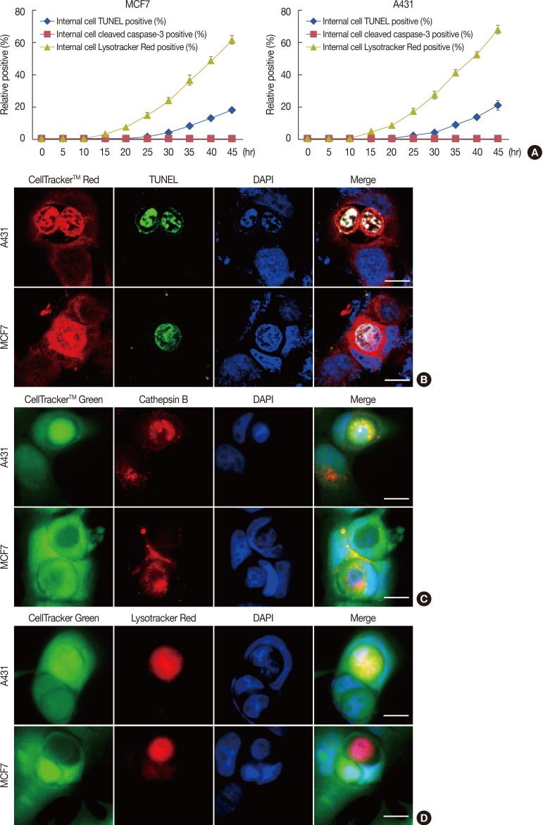
Figure 1. Lysosome-dependent cell-in-cell death of A431 and MCF-7 cell lines. (A) Kinetic quantification of internalized terminal-deoxynucleotidyl transferase mediated nick end labeling (TUNEL) positive cells, internalized cells containing cleaved caspase-3 and internalized cells demonstrating lysosome activation in cell-in-cell structures of A431 cells and MCF-7 cells. One representative experiment of three independent experiments is shown. Data are presented as means±SD. (B) Confocal images show TUNEL positive (green) and DNA fragmentation (blue) of internalized A431 cells (red) and MCF-7 cells (red) at 48 hours. Both internalized A431 and MCF-7 showed inconspicuous changes in cell size and gradual nuclei degradation after entosis. Nuclei were labeled with 4',6-diamidino-2-phenylindole (DAPI). The scale bars are 10 µm. (C) As early as 6 hours after cell engulfment, the release of active cathepsin B (red) from the lysosomes into the plasma of the internalized cells was detected and was also seen in the surrounding cytoplasm of outer cells. Cells were stained with CellTracker™ Green and cell nuclei were labeled with DAPI. The scale bars are 10 µm. (D) A431 cells (green) and MCF-7 (green) cells are stained for lysosomes using LysoTracker™ Red (red), which binds to acidified compartments after 30 hours of culture. Confocal images show positive staining for cathepsin B in the cytoplasm of internalized cells. Scale bars are 10 µm.

Figure 1