. 2016 Sep 23;19(3):308–315. doi: 10.4048/jbc.2016.19.3.308

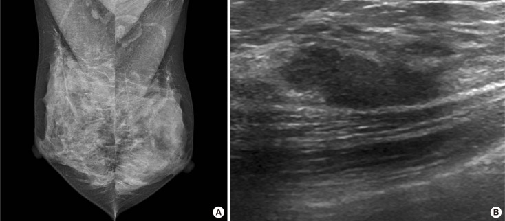
Figure 2. Radiologic findings of a 38-year-old woman with a several-week history of spontaneous serous discharge from the right nipple. (A) Mediolateral mammography of the both breast shows dense breasts with no abnormalities to explain right nipple discharge (Breast Imaging Reporting and Data System [BI-RADS] category 1). (B) Longitudinal ultrasound images show a 2.0 cm sized isoechoic, oval shaped, microlobulated mass with parallel orientation which was located at 1 o'clock 2 cm from nipple (BI-RADS category 3). Ultrasound-guided vacuum-assisted biopsy was performed and the pathologic result was consistent with fibrocystic disease with stromal fibrosis. After the procedure, the patient had no persistent nipple discharge, and no malignancy was detected during a 2-year follow-up period.

Figure 2