. 2016 Sep 23;19(3):324–329. doi: 10.4048/jbc.2016.19.3.324

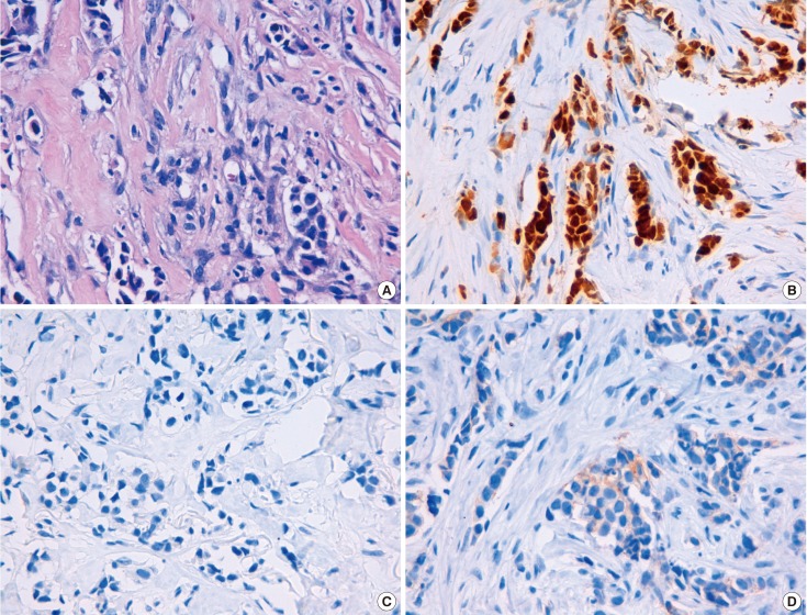
Figure 1. Pathologic findings of primary breast carcinoma. (A) Microphotograph showing invasive ductal carcinoma of the breast growing within a desmoplastic stroma (H&E stain, ×400). (B) Immunohistochemical stain showing positivity for estrogen receptor (90%) (×400). (C) Immunohistochemical stain showing absence of expression of progesterone receptor (×400). (D) Immunohistochemical stain showing negativity for human epidermal growth factor receptor 2 expression (1+) (×400). The aforementioned histologic features and the immunohistochemical expression pattern of the tumor were consistent with a diagnosis of invasive breast ductal carcinoma.

Figure 1