. 2016 Sep 23;19(3):324–329. doi: 10.4048/jbc.2016.19.3.324

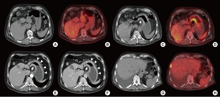
Figure 2. Pre- and postoperative positron emission tomography/computed tomography (PET/CT) finding. (A) Unenhanced CT and (B) Fusion PET/CT axial images showing no gastric fluorodeoxyglucose (FDG) uptake neither gastric wall thickening. (C, D) PET/CT showing the wall thickening of the gastric antrum (white arrow head) with a significant FDG uptake (black arrow head). (E) CT axial arterial and (F) venous phase, respectively at 35 and 80 seconds after intravenous iodinate contrast medium injection, showing the progression of the wall thickening, now involving the antrum and the body of the stomach, figuring out a linitis plastic (white arrow heads). (G, H) PET/CT axial images performed after the gastric resection showing no FDG uptake at the level of Roux-en-Y esophagojejunal anastomosis.

Figure 2