. 2016 Sep 23;19(3):330–333. doi: 10.4048/jbc.2016.19.3.330

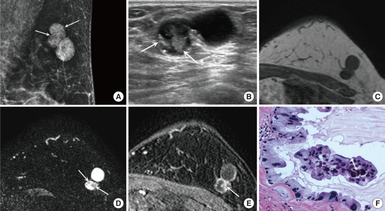
Figure 1. A 59-year-old female with mucinous cystadenocarcinoma (MCA) in the left breast. (A) Mediolateral oblique view of mammography shows a 2.8 cm sized lobular-shaped circumscribed hyperdense mass containing round or punctate microcalcifications (arrows) in the upper outer quadrant of the left breast. (B) On ultrasound, the MCA appears as a circumscribed complex cystic and solid mass. The solid portion of the mass (arrows) shows an irregular shape and isoechogenicity, with multiple echogenic dots corresponding to the microcalcifications seen on mammography. On magnetic resonance imaging, the solid portion shows low signal intensity (SI) on T1 weighted imaging (WI) (C) and intermediate SI on T2-WI (arrows) (D). The cystic portion shows rim-enhancement and the solid portion (arrow) shows nodular enhancement on dynamic contrast-enhanced image with persistent enhancement kinetics (E). (F) The tumor is composed of multiple cysts distended by mucinous secretion and the cysts are lined with tall columnar cells with intracytoplasmic Most nuclei are basally located and somewhat bland, however some nuclei show more atypia. Note the psammomatous calcification (arrow) in the papillary core (H&E stain, ×400).

Figure 1