. 2016 Sep 23;19(3):330–333. doi: 10.4048/jbc.2016.19.3.330

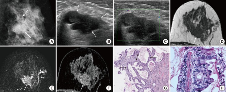
Figure 2. A 50-year-old female with mucinous cystadenocarcinoma in the left breast. (A) Mediolateral oblique view of mammography shows a 2.8 cm sized lobular-shaped isodense mass with obscured margin and a few faint microcalcifications in the left mid outer breast (arrows). (B) A multilobulated complex cystic and solid mass is seen on ultrasound (US). The solid portion of the mass (arrows) is hypoechoic and the margin is partially indistinct. (C) Vascularity is not increased and a vascular stalk is not detected on Doppler US. On magnetic resonance imaging, a cystic and solid mass is shown. The solid portion (arrow) has an irregular shape and the margin shows hypointensity on T1 weighted imaging (WI) (D), intermediate signal intensity on T2-WI (E), and heterogeneous enhancement with persistent enhancement kinetics (F). (G) Core biopsy shows multilocular cystic lesion containing extracellular mucin. The cyst wall is lined with tall columnar mucinous epithelium (arrows) (H&E stain, ×100). (H) Mucinous epithelium shows marked atypia and pleomorphism. There are frequent mitotic figures (H&E stain, ×400).

Figure 2