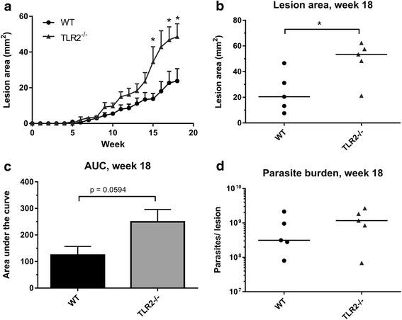
Fig. 3.

Infection of WT and TLR2−/− mice with L. mexicana LPG1 −/− promastigote parasites. WT and TLR2−/− mice were infected with 105 L. mexicana LPG1 −/− parasites, and the disease was monitored by measuring the lesions every week for 18 weeks (n = 5). The average lesion size (mm2) + standard error (SEM) are displayed for both groups at all time points post-infection (a), and at the end of the experiment (week 18, b). The AUC was calculated for each mouse after the 18 weeks, the average is displayed (+SEM) in the bar chart in (c). The parasite burden in the lesion tissue was determined by qPCR, and individual burdens and median averages are displayed in (d). Groups were compared using a Mann-Whitney U test where P < 0.05 was considered to indicate significant (*) differences
