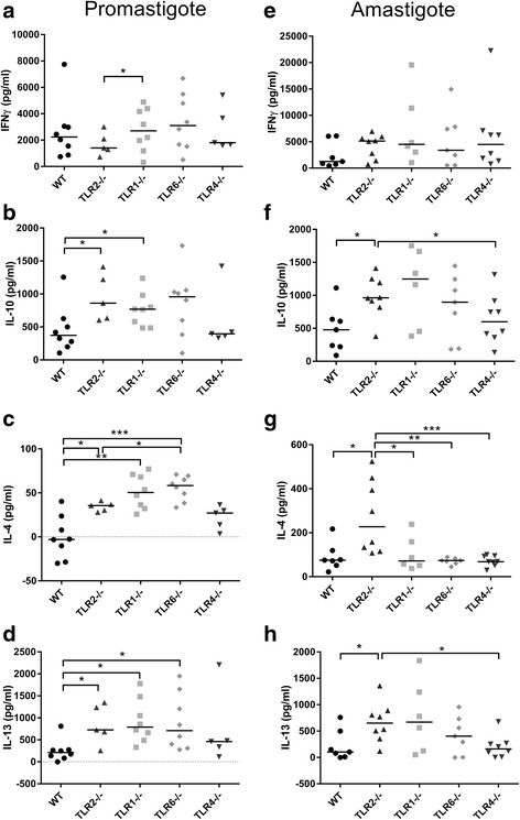
Fig. 5.

Antigen-specific cytokine production in L. mexicana infected WT and TLR−/− mice. WT, TLR2−/−, TLR1−/−, TLR6−/− and TLR4−/− mice were infected with L. mexicana parasites (promastigotes a, b, c, d, amastigotes e, f, g, h) and left to develop lesions for 14 weeks. At the end of the experiment, DLN were removed and the cells were re-stimulated for 72 h in vitro with the Leishmania antigen FTAg. The supernatants were collected and analysed for the presence of the cytokines IFNγ, IL-10, IL-4 and IL-13 using ELISA. The quantities of cytokine produced in response to FTAg are shown for each individual, along with the median values for each group. Groups were compared using a Mann-Whitney U test where P < 0.05 (*) and P < 0.01 (**) were considered to indicate significant differences
