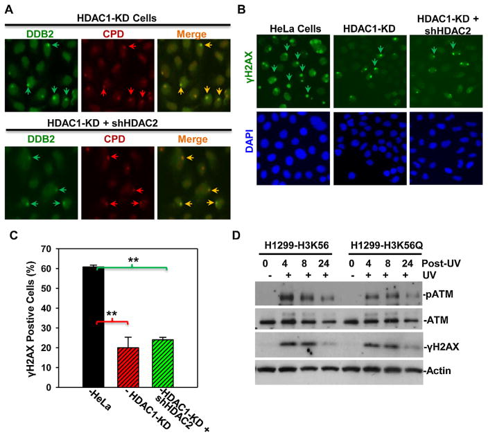
Fig. 5.
(A) HDAC1/2 knockdown does not affect DDB2 recruitment to DNA damage sites. (A) Stable HeLa HDAC1-KD cells or HDAC1-KD cells transiently transfected with shHDAC2 DNA constructs were UV irradiated at 100 J/m2 through a 5-μm micropore filter and maintained for 1 h. The cells were then fixed with 2% paraformaldehyde, and, DDB2 and CPD were visualized by immunofluorescence using anti-DDB2 or anti-CPD specific antibodies. Arrows indicated typical UVR-induced, antibody-decorated immunofluorescence spots of DDB2 and CPD. (B) HDAC1 and HDAC2 knockdown decreases γH2AX accumulation at DNA damage after UV irradiation. Stable HeLa HDAC1-KD cells or HDAC1-KD cells transiently transfected with shHDAC2 DNA constructs were UV irradiated at 20 J/m2 through a 5-μm micropore filter. The cells were fixed with 2% paraformaldehyde 1 h after UV irradiation, and, γH2AX foci (pointed by arrows) were visualized at DNA damage spots by immunofluorescence. (C) Quantitative data are from the analysis of γH2AX foci in HeLa, HeLa HDAC1-KD cells and hHDAC2-tranfected HDAC1-KD cells. Mean ± SE were calculated from multiple microscopic fields of three independent experiments. Double asterisk mark (**) indicates p ≤ 0.01. (D) H1299 cells expressing H3K56 or H3K56Q were irradiated at 20 J/m2 and harvested at indicated time points. ATM, phospho-ATM and γH2AX were determined by specific antibodies. Actin blots were used as loading control.

