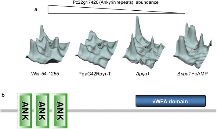
Fig. 6.

a Abundance of the Pc22g17420 protein in the different strains/conditions. The fluorescence signal was obtained with the program DeCyder 2-D differential analysis software. b Schematic representation of the Pc22g17420 protein, showing the ankyrin repeat domain close to the N-terminal end and the vWFA domain at the C-terminal region. The domain structure of the protein was generated with SMART (simple modular architecture research tool, http://smart.embl-heidelberg.de)
