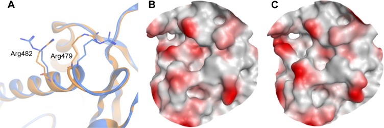
Figure 5.
The active site of two crystal structures of CDC25B aligned on each other.
Notes: (A) PDB: 1CWR40 is the apo structure and shown in gold cartoon, whereas 1QB040 is the ligand-bound structure and shown in blue cartoon. (B) The binding pocket surface of the apo structure of CDC25B. (C) The binding pocket surface of the CDC25B ligand-bound structure.

