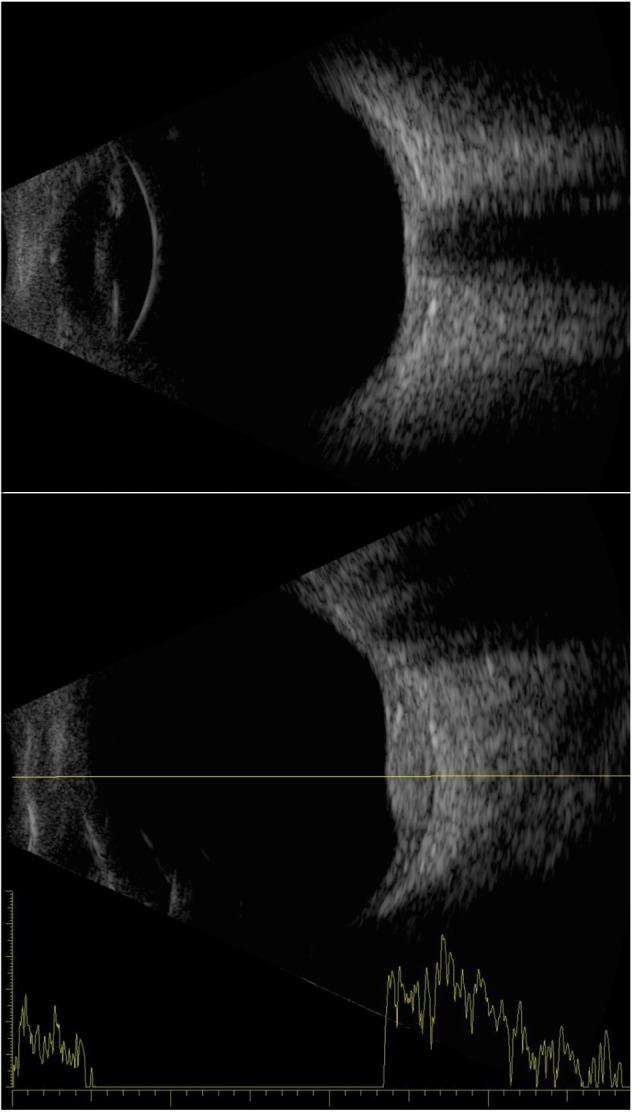
Figure 3.

Examples of an axial B-scan of a normal eye (top) and a combined B-/A-scan of an eye with a choroidal hemangioma temporal to the disc (bottom).
Notes: The axial contact B-scan demonstrates the anterior chamber, iris/pupil, lens, and posterior pole, including the optic nerve. The hemangioma demonstrates sustained high-amplitude echoes on A-scan.
