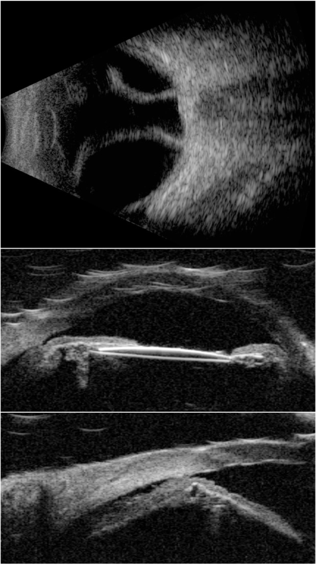
Figure 5.

A 10 MHz B-scan (top) and UBM images (center and bottom) of an eye with choroidal detachment and ciliary body effusion over 360° following cataract surgery with IOL implantation.
Note: IOL is seen in the center image.
Abbreviations: IOL, intraocular lens; UBM, ultrasound biomicroscopy.
