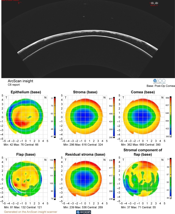
Figure 6.
Top: insight-100 arc scan image of a 1-year post-LASIK cornea and pachymetric maps (bottom) derived from a series of scans along each clock-hour.
Notes: Maps represent (top row, left to right) epithelial, stromal, and corneal thickness and (bottom row, left to right) flap depth, residual stromal thickness, and stromal component of flap thickness. Image courtesy Prof Dan Z. Reinstein, MD MA (Cantab) FRCSC DABO FRCOphth FEBO.
Abbreviations: LASIK, laser-assisted in situ keratomileusis; min, minimum; max, maximum.

