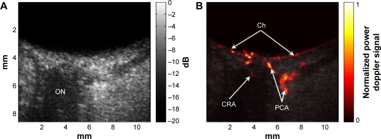
Figure 8.
Ultrafast plane-wave angiographic imaging of the posterior pole.
Notes: B-scan (A) and high-resolution blood-flow image (B) of the posterior pole of a normal eye derived from coherently compounded plane-wave images (20 angles over ±10°) with data acquired continuously for 1 s at 20,000 planes/s (1,000 compound images/s) with an 18 MHz linear array.
Abbreviations: Ch, choroid; CRA, central retinal artery; ON, optic nerve; PCA, posterior ciliary arteries; s, seconds.

