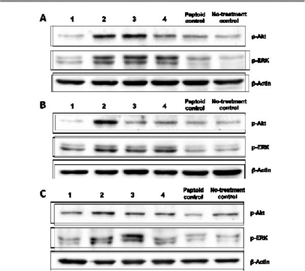
Fig. 7.
Effects of hit peptoids 1-4 on the expression levels of p-Akt and p-ERK in (A) MCF-7, (B) MDA-231, and (C) NIH-3T3 cells. After serum starvation , all of the three cell lines were treated with peptoids 1-4 and a randomly selected peptoid control at a concentration of 15 μM for 10 min. The expression levels of p-Akt and p-ERK were detected by Western blot using specific antibodies.

