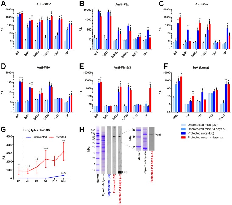
Fig 7. Serum and pulmonary antibody profiles in unprotected and protected mice following B. pertussis challenge.
Serum IgA, IgG, and IgG subclass responses specific for (A) OMV, (B) Ptx, (C) Prn, (D) FHA, and (E) Fim2/3 were determined by using a MIA. Data were obtained in naive mice and protected mice prior to challenge (D0) and 14 days post infection (p.i.) or 14 days post-challenge (p.c.) (n = 3/time point). (F) Pulmonary IgA responses against these antigens were determined on the same time points. * = p<0.05 experimental group versus unprotected group (D0), + = p<0.05 protected group (D0) versus protected group (14 days p.c.). (G) The kinetics of the anti-OMV IgA antibody formation in lung lysates were analyzed at more time points (n = 3/time point) and expressed in fluorescence intensity (F.I.). **** = p<0.0001, challenged unprotected or protected group versus unprotected or protected group (day 0), ++ and +++ = p<0.01 and p<0.001 unprotected group versus protected group (for each time point). (H) Western blot on separated B. pertussis B1917 proteins was performed with pooled lung lysates (1:50) of unprotected and protected mice prior to challenge (D0), and of protected mice 14 days p.c. with IR800-labeled secondary antibody. Left panel shows whole protein range (260kDa-<10kDa) of B. pertussis lysate. Right panel shows more detailed separation of the 110-60kDa protein range. Antigen identification for Vag8 and LPS is depicted.

