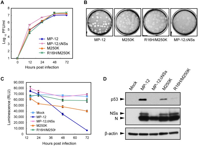
Fig 3. Attenuated cytotoxicity of MP-12 NSs mutants.
(A) Growth kinetics of MP-12 and its mutants. Vero E6 cells were infected with each virus at an m.o.i. of 0.01, and the culture supernatants were collected at various times p.i. Virus titers were determined by plaque assays. (B) Plaque morphology of the MP-12 and mutant viruses in VeroE6 cells at 3 day p.i. (C) Viability of cells infected with MP-12 or its mutants infected cells. Vero E6 cells were mock infected or infected with each virus at an m.o.i. of 3. Cell viability was determined by measuring cellular ATP. (D) Abundance of p53 in infected cells. Vero E6 cells were infected with each virus at an m.o.i. of 3 and harvested at 24 h p.i. Whole cell lysate was subjected to Western blot analysis. Anti-p53 antibody, anti-MP-12 antibody and anti-β-actin antibody were used as the primary antibody for the top, middle and bottom panels, respectively.

